Sunday, May 7, 2017

The Shape of a Virus

Viruses are so small of a size (20-750 nm) that most can only be seen through an scanning electron microscope.
Structure of a virus:

  • genetic information molecule
  • protein layer that envelopes the above molecule (capsid)
  • Inside there are nucleic acids that form either RNA/DNA.
There are 4 known virus shapes:

  1. Helical
  2. Icosahedral 
  3. Envelope
  4. or a combination called complex
------------------------------------------------------------------------------------------------------------The helical shaped virus has protein organized in a circular path. This forms a tube with a hollow core that holds nucleic acid. 
Width: 15-19 nm
Length: 300-500 nm
virus known to have this shape: filamentous virus ex. tobacco mosaic virus
filamentous virus usually infect gram negative bacteria.

------------------------------------------------------------------------------------------
The icosahedral shaped virus has a sphere-like shaped made from fused equilateral triangles. These triangles are "identical protein sub units". This prime shape holds the genetic material within. Virus's with this shape tend to be released when a cell lyses. 
amount of protein sub-units: 20 faces
size: 70-90 nm
know viruses with this shape: polio virus, rhinovirus, and adenovirus


---------------------------------------------------------------------------------------------
The envelope shaped virus is a standard helical/isocahedral virus except it is encased in a lipid bilayer membrane.These membranes come from the host cell. 
known viruses of this type: hepatitis C, HIV, and influenza virus.
------------------------------------------------------------------------------------------------------------
Complex type of virus have a head and tail they also only infect bacteria. The tail creates a hole in cell wall of caught bacteria then releases DNA into it. These complex viruses are among the largest. Also known as bacteriophage
Examples of this type: smallpox virus(variola).

========================================================================

https://youtu.be/CBMr142ynbk
https://youtu.be/Ofd_lgEymto
https://dr282zn36sxxg.cloudfront.net/datastreams/f-d%3A0af319c1f63d9f6a49d9b6245463fc8fea256fd364c6a8e427999d1d%2BIMAGE_THUMB_POSTCARD_TINY%2BIMAGE_THUMB_POSTCARD_TINY.1
http://figures.boundless-cdn.com/14011/raw/tevenphage.svg
https://upload.wikimedia.org/wikipedia/commons/f/f0/Lipid_bilayer_section.gif
https://youtu.be/_HvKxgihn4I
http://images.slideplayer.com/34/8309974/slides/slide_6.jpg
https://morgridge.org/wp-content/uploads/Virus-Structure.pdf
http://www.twiv.tv/wp-content/uploads/2009/07/helical-symmetry2-1024x775.jpg

Reproductive System

Female Reproductive System: Consists of the ovaries, vagina, uterus, Fallopian tubes, breasts, and mammary glands. Their function is to produce gametes then transport them, and also make sex hormones. Among other functions this system also allows for the ova to be  fertilized by sperm, and aide in the growth of offspring. The system will create ovum in order for the uterus to be prepared if pregnancy is to occur. This is called the reproductive cycle which occurs for about 28 days on average, more or less for some woman. If the ovum is not fertilized it will be menstruated. The luteinizing hormone and follicle stimulating hormone will urge the ovaries to make a developed ovum which is called ovulation, and happens after 14 days into the reproductive cycle. Each ovum begins as an oocyte before it reaches maturity. The ovum then goes through a week long process to the Fallopian tubes then finally to the uterus. once fertilized the ovum can become a zygote. After thorough cell division the zygote will become an embryo, which then embeds into the uterine wall. The uterine wall is known as the endometrium. If the ovum does not get fertilized the arteries in the uterus halt blood flow to the endometrium. The dead cells are then shed away in a process called menstruation.
Male Reproductive System: The penis, spermatic ducts, sex glands, testes, and scrotum make up this system. All together these parts will produce sperm, male gamete to create the semen. Semen which will be responsible to fertilize the ovum. LH and FSH prepare the body for spermatogenesis at the start of puberty. LH starts off testosterone in the testes. FSH will allow germ cells to develop fully. When fertilizing an egg sperm will use special enzyme to get through the corona radiata, and zona pellucida layers of the egg.

Reproductive: relating to or effecting reproduction.
cervix: the narrow neck ike passage forming the lower end of the uterus.
clitoris: a small sensitive and erectile part of the female genitals at the anterior end of the vulva.
Contraception: the deliberate use of artificial methods or other techniques to prevent pregnancy as a consequence of sexual intercourse. 
corpus luteum: a hormone-secreting structure that develops in an ovary after an ovum has been discharged but degenerates after a few days unless pregnancy has begun.
Cowper’s gland: either of a pair of small glands that open into the urethra at the base of the penis and secrete a constituent of seminal fluid.
ejaculatory duct: about two centimeters in length and is created when the seminal vesicle's duct merges with the vas deferens.
epididymis: a highly convoluted duct behind the testis, along which sperm passes to the vas deferens.
Estrogen: any of a group of steroid hormones that promote the development and maintenance of female characteristics of the body. 
Follicles: a small secretory cavity, sac, or gland, in particular.
FSH:  a gonadotropin, a glycoprotein polypeptide hormone. FSH is synthesized and secreted by the gonadotropic cells of the anterior pituitary gland, and regulates the development, growth, pubertal maturation, and reproductive processes of the body.
GnRH: gonadotropin-releasing hormone. This hormone is released by the hypothalamus in the brain.GnRH acts on receptors in the anterior pituitary gland.
LH: a hormone produced by gonadotropic cells in the anterior pituitary gland.
menopause: the ceasing of menstruation.
Ovaries: a female reproductive organ in which ova or eggs are produced, present in humans and other vertebrates as a pair.
Ovulation: discharge of ova or ovules from the ovary.
Penis: the male genital organ of higher vertebrates, carrying the duct for the transfer of sperm during copulation. In humans and most other mammals, it consists largely of erectile tissue and serves also for the elimination of urine.
Progesterone: a steroid hormone released by the corpus luteum that stimulates the uterus to prepare for pregnancy.
prostate gland: a gland surrounding the neck of the bladder in male mammals and releasing prostatic fluid.
scrotum: a pouch of skin containing the testicles.
semen: the male reproductive fluid, containing spermatozoa in suspension.
seminal vesicle: each of a pair of glands that open into the vas deferens near its junction with the urethra and secrete many of the components of semen.
sperm: the mature motile male sex cell of an animal, by which the ovum is fertilized, typically having a compact head and one or more long flagella for swimming.
testis: an organ that produces spermatozoa 
Testosterone: a steroid hormone that stimulates development of male secondary sexual characteristics, produced mainly in the testes, but also in the ovaries and adrenal cortex.
Uterus:the organ in the lower body of a woman or female mammal where offspring are conceived and in which they gestate before birth; the womb.
vagina: the muscular tube leading from the external genitals to the cervix of the uterus in women and most female mammals.
Vas deferens: the duct that conveys sperm from the testicle to the urethra.
Vestibule: a chamber or channel communicating with or opening into another.
vulva: the female external genitals.

http://stream1.gifsoup.com/view1/20150310/5183019/fertilization-o.gif
https://media1.britannica.com/eb-media/31/94931-004-4DED6AD1.jpg
http://www.innerbody.com/image/repfov.html#full-description

Urinary System


Urethra, ureter, urinary bladder, and kidneys are the urinary system. This system exist to eliminate waste, and let out urine from the body. The urinary tract gets urine from the kidneys which then keeps it, and later releases it. The system also works to help maintain homeostasis of water, pH, blood pressure, red blood cells, calcium levels, and ions all intact. Magnesium, sodium, calcium, chloride, and potassium ions are secreted into urine from the kidneys. Kidneys also control levels of bicarbonate, and hydrogen ions. The hydrogen ions yield a surplus of H+ which the kidneys must excrete. Bicarbonate ions form the natural buffer in the blood. Nephrons in the kidney transform blood to urine. Renin, Calcitriol, and erythropoietin are hormones that the kidney produces that interact with other systems in the body.
Arteriole: small artery
calyx or calix: Cup-like collecting region of the renal pelvis (plural: calyces or calices)
Catheter: Tube for injecting or removing fluids
Cortex: outer region of an organ;the renal cortex is the outer region of the kidney (cortical means pertaining to the cortex)
Creatine: Nitrogenous waste excreted in urine. Creatinine clearance is a measure of the efficiency of the kidneys in removing (clearing) creatinine from the blood
Electrolyte: chemical element that carries an electrical charge when dissolved in water. Electrolytes are necessary for functioning of muscles and nerves. The kidneys maintain the proper balance of electrolytes and water in the blood. Potassium (K+) and Sodium (Na+) are electrolytes
erythropoietin (EPO): Hormone secreted by the kidney to stimulate the production of red blood cells by bone marrow. --poietin means a substance that forms.
Filtration: process whereby some substances, but not all, pass through a filter. In the kidney, blood pressure forces materials through the filter (glomerulus). About 180 quarts of fluid are filtered from the blood daily, but the kidney returns 98% to 99% of the water and salts. Only about 1.5 quarts (1500 mL) of urine are excreted daily
glomerular capsule: Enclosing structure surrounding each glomerulus. The glomerular capsule is known as Bowman's capsule and it collects the material that is filtered from the blood through the walls of the glomerulus
Glomerulus: Tiny ball of capillaries (microscopic blood vessels) in the kidney. Plural: glomeruli
Hilum: Depression in an organ where blood vessels and enter and leave.
Kidney: One of two bean-shaped organs on either side of the backbone in the lumbar region; it filters nitrogenous wastes from the bloodstream to form urine.
Meatus: opening or canal
Medulla: inner region of an organ. The renal medulla is the inner region of the kidney (medullary means pertaining to the medulla)
Nephron: Combination of glomerulus and renal tubule where filtration, re-absorption , and secretion take place in the kidney. It is the functional unit of the kidney; each capable of forming urine by itself. There are about 1 million nephrons in a kidney
nitrogenous waste: substance containing nitrogen and excreted in urine.
Potassium: An electrolyte regulated by the kidney so that a proper concentration is maintained within the blood. potassium is essential for allowing muscle contraction and conduction of nervous impulses. (K+)
Reabsorption: process whereby renal tubules return materials necessary to the body back into the bloodstream
renal artery: Blood vessel that carries blood to the kidney
renal pelvis: Central collection region in the kidney
renal tubules: microscopic tubes in the kidney where urine is formed after filtration
renal vein: blood vessel that carries blood away from the kidney and toward the heart
Renin: hormone secreted by the kidney; it raises blood pressure by influencing vasoconstriction (narrowing of blood vessels)
Sodium: An electrolyte regulated in the blood and urine by the kidneys; needed for proper transmission of nerve impulses, heart activity, and other metabolic functions
Trigone: Triangular area in the urinary bladder.
Urea: major nitrogenous waste excreted in urine.
Ureter: One of two tubes leading from the kidneys to the urinary bladder.
Urethra: Tube leading from the urinary bladder to the outside of the body.
uric acid: nitrogenous waste excreted in the urine
urinary bladder: hollow, muscular sac that holds and stores urine.
Urination: process of expelling urine; also called micturition (voiding)
http://stream1.gifsoup.com/view4/4688965/urinary-system-o.gif
http://antranik.org/wp-content/uploads/2011/12/organs-of-urinary-system-cadaverrenal-artery-renal-hilum-renal-vein-1024x558.jpg

Digestive System


This system is made of the gall bladder, pancreas, liver, large/small intestine, stomach, esophagus, and mouth. Everything listed except the gallbladder, and pancreas are called the gastrointestinal tract. Besides these parts there are also gut flora which are bacteria that control part of digestion. Nutrients that the body needs for growth, healing of cells, and energy are synthesized from the breakdown of food. All of these nutrients can be grouped into one of the four categories which are fats, proteins, carbohydrates, and vitamins. The mouth chews food and saliva is the digestive juice that breaks it down to a starch. The esophagus then brings down the food by swallowing to the stomach. From then the muscles calms down to allow food and stomach acid breaks it down further to carbohydrates, proteins, and starches. The mixture from the stomach called chyme goes to the small intestine which gives nutrients to the blood stream. From there large intestine collects leftover nutrients and water then forms stool for bowel movement. Pancreatic juice is also secreted to form starches, fats, and proteins. The liver produces bile acid to create fats.  
alimentary canal: the whole passage along which food passes through the body from mouth to anus. It includes the esophagus, stomach, and intestines.
Amylase: an enzyme, found chiefly in saliva and pancreatic fluid, that converts starch and glycogen into simple sugars.
Bile: a bitter greenish-brown alkaline fluid that aids digestion and is secreted by the liver and stored in the gallbladder.
Chyme: the pulpy acidic fluid that passes from the stomach to the small intestine, consisting of gastric juices and partly digested food.
Digestion: the process of breaking down food by mechanical and enzymatic action in the alimentary canal into substances that can be used by the body.
Duodenum: the first part of the small intestine immediately beyond the stomach, leading to the jejunum.
Epiglottis: a flap of cartilage at the root of the tongue, which is depressed during swallowing to cover the opening of the windpipe.
esophagus: the part of the alimentary canal that connects the throat to the stomach; the gullet. In humans and other vertebrates it is a muscular tube lined with mucous membrane.
Feces: waste matter discharged from the bowels after food has been digested; excrement.
gall bladder: the small sac-shaped organ beneath the liver, in which bile is stored after secretion by the liver and before release into the intestine.
gastric juice: a thin, clear, virtually colorless acidic fluid secreted by the stomach glands and active in promoting digestion.
HCl: hydrogen chloride
large intestine: the cecum, colon, and rectum collectively.
Liver: a large lobed glandular organ in the abdomen of vertebrates, involved in many metabolic processes.
mesentery: a fold of the peritoneum that attaches the stomach, small intestine, pancreas, spleen, and other organs to the posterior wall of the abdomen.
Mucus: a slimy substance, typically not miscible with water, secreted by mucous membranes and glands for lubrication, protection
Pancreas: a large gland behind the stomach that secretes digestive enzymes into the duodenum. Embedded in the pancreas are the islets of Langerhans, which secrete into the blood the hormones insulin and glucagon.
pancreatic juice:  the clear alkaline digestive fluid secreted by the pancreas.
Pepsin: the chief digestive enzyme in the stomach, which breaks down proteins into polypeptides.
pepsinogen: a substance that is secreted by the stomach wall and converted into the enzyme pepsin by gastric acid.
Peristalsis: the involuntary constriction and relaxation of the muscles of the intestine or another canal, creating wavelike movements that push the contents of the canal forward.
pH: a measure of hydrogen ion concentration; a measure of the acidity or alkalinity of a solution.
Pharynx: the membrane-lined cavity behind the nose and mouth, connecting them to the esophagus.
pyloric sphincter: a band of smooth muscle at the junction between the pylorus of the stomach and the duodenum of the small intestine.
Rectum: final section of the large intestine, terminating at the anus.
small intestine: the part of the intestine that runs between the stomach and the large intestine; the duodenum, jejunum, and ileum collectively.
Stomach:  internal organ in which the major part of the digestion of food occurs, being (in humans and many mammals) a pear-shaped enlargement of the alimentary canal linking the esophagus to the small intestine.
Villi: any of numerous minute elongated projections set closely together on a surface, typically increasing its surface area for the absorption of substances, in particular.
http://i.imgur.com/BCmo8eX.gif
http://www.digestivesystem.organsofthebody.com/images/digestive-system-facts-thumb.jpg

Respiratory System


Parts of this system include lungs, diaphragm, intercostal muscles, mouth, nose, larynx, pharynx, trachea, and bronchi which are considered the airway. These work together to provide oxygen for the cells. This system must also dispose of CO2. Alveoli and atmosphere produce a negative pressure gradient. The lungs closed off by the pleural membrane create a pressure that is less than that of the atmosphere’s in a resting state. Which leads air to fill the lungs when at rest. Air fills the lungs until the pressure is equal to that of the atmosphere. The diaphragm, and external intercostal muscles contracts in response allowing more air to flow in. The thorax volume increases, and the pressure of the lungs goes below that of the atmosphere. When air is exhaled the external intercostal muscles, and diaphragm must relax. The internal intercostal muscles contract. The thorax in turn decrease in volume, and then increases pressure of the lungs. The switched direction of the pressure gradient allows air to leave until air pressures match inside and out of the body.

alveolar duct: The part of the respiratory passages beyond the respiratory bronchioles, from which the alveolar sacs and alveoli arise.
Alveoli: tiny air sacs of the lungs which allow for rapid gaseous exchange.
bicarbonate ions: a salt of carbonic acid containing the ion HCO3--; an acid carbonate.
bronchial tree: the branching system of bronchi and bronchioles conducting air from the windpipe into the lungs.
Bronchiole: branches into which a bronchus divides.
Cilia: a short, microscopic, hairlike vibrating structure.
diaphragm: a dome-shaped, muscular partition separating the thorax from the abdomen in mammals. It plays a major role in breathing, as its contraction increases the volume of the thorax and so inflates the lungs.
Expiration: exhalation of breath.
gas exchange: The diffusion of gases from an area of higher concentration to an area of lower concentration
hemoglobin: a red protein responsible for transporting oxygen in the blood of vertebrates. Its molecule comprises four subunits, each containing an iron atom bound to a heme group.
inspiration: the drawing in of breath
larynx: the hollow muscular organ forming an air passage to the lungs and holding the vocal cords in humans and other mammals; the voice box.
Lung: each of the pair of organs situated within the rib cage, consisting of elastic sacs with branching passages into which air is drawn, so that oxygen can pass into the blood and carbon dioxide be removed
Pharynx: the membrane-lined cavity behind the nose and mouth, connecting them to the esophagus.
pleural cavity: the space that lies between the pleura, the two thin membranes that line and surround the lungs.
Respiration: the action of breathing.
respiratory control center: located in the medulla oblongata and pons, which are parts of the brainstem. The RCs receive controlling signals of neural, chemical, and hormonal nature and control the rate and depth of respiratory movements of the diaphragm and other respiratory muscles.
Trachea: a large membranous tube reinforced by rings of cartilage, extending from the larynx to the bronchial tubes and conveying air to and from the lungs; the windpipe.
Image result for respiratory system gif
https://www.google.com/url?sa=i&rct=j&q=&esrc=s&source=images&cd=&cad=rja&uact=8&ved=0ahUKEwiduruR-97TAhUIS2MKHf4gAdQQjhwIBQ&url=https%3A%2F%2Fwww.docsity.com%2Fen%2Fnews%2Feducation-2%2Fsystems-human-body-interactive-gifs%2F&psig=AFQjCNHzCDhpHdzgG1QMtzrw4kZqAWq0Zw&ust=1494286976703929
http://www.therespiratorysystem.com/wp-content/themes/Respiratory/images/respiratory-system-diagram.jpg

Cardiovascular System


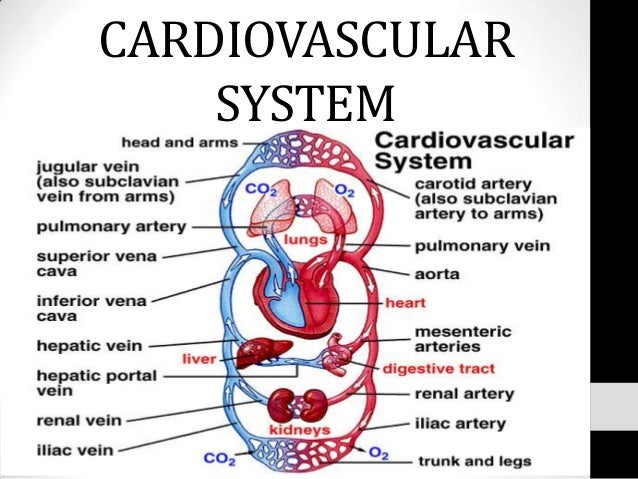
This system is made up of the heart, veins, capillaries, and arteries (blood vessels). The purpose of this system is to transfer oxygen, and nutrients to tissues, and then get the waste products to the organs that can properly dispose of them. Veins bring blood into the heart while arteries carry the blood from the heart around the whole body. The body’s total supply of blood is at around 5 liters. Which the heart is able to pump out every minute. There are two types of circulatory loops the pulmonary and systemic circulation loops. Blood vessels are lined with simple squamous epithelium which has this to stop clots, and keep blood inside. Inside the heart epithelium is called the endocardium. This system transports, maintains homeostasis, and protects. To regulate the blood vessels open up to spill out blood during times of heat, and when it is cold they constrict to keep blood flowing to the core. The blood vessels also help to make a buffer to keep the pH stable, and they create an isotonic environment for cells. The white blood cells serve to fight pathogens, and clean cellular waste, the red blood cells get with platelets to prevent blood loss, and cover open wound. Antibodies are stored in blood to protect from invaders, and hold immunity to previous pathogens.
Antibodies: a blood protein produced in response to and counteracting a specific antigen. Antibodies combine chemically with substances that the body recognizes as alien, such as bacteria, viruses, and foreign substances in the blood.
antigens: a toxin or other foreign substance that induces an immune response in the body, especially the production of antibodies.
Aorta: the main artery of the body, supplying oxygenated blood to the circulatory system. In humans it passes over the heart from the left ventricle and runs down in front of the backbone.
Arteriole: a small branch of an artery leading into capillaries.
artery: any of the muscular-walled tubes forming part of the circulation system by which blood (mainly that which has been oxygenated) is conveyed from the heart to all parts of the body.
Atrium: each of the two upper cavities of the heart from which blood is passed to the ventricles. The right atrium receives deoxygenated blood from the veins of the body; the left atrium receives oxygenated blood from the pulmonary vein.
Capillary: any of the fine branching blood vessels that form a network between the arterioles and venules.
circulatory system: the system that circulates blood and lymph through the body, consisting of the heart, blood vessels, blood, lymph, and the lymphatic vessels and glands.
diastole: the phase of the heartbeat when the heart muscle relaxes and allows the chambers to fill with blood.
ECG: a simple, noninvasive procedure.
Myocardium: the muscular tissue of the heart.
Pacemaker: an artificial device for stimulating the heart muscle and regulating its contractions.
peripheral resistance: near the surface of the body, with special reference to the circulation and nervous system.
Platelets: a small colorless disk-shaped cell fragment without a nucleus, found in large numbers in blood and involved in clotting.
pulmonary circulation: is the portion of the cardiovascular system which carries deoxygenated blood away from the heart, to the lungs, and returns oxygenated (oxygen-rich) blood back to the heart.
Purkinje fibers: carry the contraction impulse from both the left and right bundle branch to the myocardium of the ventricles.
red blood cells: typically a biconcave disc without a nucleus. Erythrocytes contain the pigment hemoglobin, which imparts the red color to blood, and transport oxygen and carbon dioxide to and from the tissues.
sinoatrial node: a small body of specialized muscle tissue in the wall of the right atrium of the heart that acts as a pacemaker by producing a contractile signal at regular intervals.
systemic circulation: part of the cardiovascular system which carries oxygenated blood away from the heart to the body, and returns deoxygenated blood back to the heart.
Systole: the phase of the heartbeat when the heart muscle contracts and pumps blood from the chambers into the arteries.
valves of the heart: any of the valves that control blood flow to and from the heart and that include the atrioventricular valves, the aortic valve, and the pulmonary valve—called also cardiac valve.
Vasoconstriction: the constriction of blood vessels, which increases blood pressure.
vasodilatation: the dilatation of blood vessels, which decreases blood pressure.
Vein: any of the tubes forming part of the blood circulation system of the body, carrying in most cases oxygen-depleted blood toward the heart.
vena cava: a large vein carrying deoxygenated blood into the heart.
Venule: a very small vein, especially one collecting blood from the capillaries.
Viscosity: the thickness and stickiness of blood.
white blood cells: a colorless cell that circulates in the blood and body fluids and is involved in counteracting foreign substances and disease; a white (blood) cell.
Image result for cardiovascular system gif
https://www.docsity.com/en/news/education-2/systems-human-body-interactive-gifs/
https://image.slidesharecdn.com/cardiovascularsystem-130721124922-phpapp01/95/cardiovascular-system-1-638.jpg?cb=1374411022